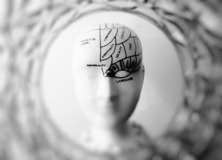
Many things can increase arousal including high temperatures, crowding, pain, loud noises, violent movies and bad odors. Arousal can also be sexually stimulating and even produce orgasms.
Arousal is important for regulating consciousness, alertness and motivation. According to Yerkes and Dodson, an optimal level of arousal improves performance on easy tasks but impairs performance on difficult ones.
The Hypothalamus
Your thalamus is a part of your brain that is responsible for interpreting sensory impulses and sending them to other parts of the brain for further processing. It also plays a role in thinking (cognition), emotions and memory. The hypothalamus, on the other hand, is responsible for the regulation of endocrine functions and is involved in sexual arousal, sleep and wakefulness, hunger, energy balance and reward.
During the first stage of sexual arousal, your heart rate and breathing increase. This is caused by signals that are relayed from the lateral hypothalamic area (LHA) to the brainstem via the locus coeruleus and the periaqueductal gray areas.
These nerve fibers are involved in the production and release of hormones, including epinephrine and norepinephrine. Epinephrine is released from the adrenal medulla and is a chemical messenger that affects the body and brain by increasing blood flow, alertness and the response to stressors. Norepinephrine, on the other hand, is a neurotransmitter that acts as a stress hormone and regulates your metabolic processes, blood pressure, and cardiovascular system.
One of the most fascinating discoveries was made when researchers found that when a woman reached the point of orgasm, her brain activity became much quieter. This is due to a reduction in activity in the dorsomedial prefrontal cortex, which controls self-control over basic desires such as sex.
The Pituitary Gland
The pituitary gland is the master endocrine gland that secretes a number of hormones involved in many body functions. Hormones are chemical messengers that are produced by neuroendocrine cells in the hypothalamus and pass to the pituitary via a series of special blood vessels called the pituitary portal system (from Latin upo phuein, meaning ‘to grow under’). The hypothalamus then signals specific cells of the pituitary that are to discharge their hormone into the blood stream. Each of these glands, known as adrenocorticotropic hormone (ACTH) cells, receive their individual’message’ from the hypothalamus and respond by releasing their own hormone into the bloodstream.
The posterior pituitary consists of two pairs of large clusters of nerve cell bodies, called nuclei, that run along the side of each ventricle of the brain and extend into the lateral wall of the third ventricle and the front part of a circular ring of blood vessels, called the circle of Willis. The’messages’ from the hypothalamus are transmitted by neuroendocrine axons that terminate in these nuclei and connect with the axons of the anterior pituitary, which is connected to the rest of the brain by a series of neural pathways that carry a variety of sensory signals including osmolality (salt concentration in fluids), thirst and circadian rhythms.
The posterior pituitary contains several types of sex hormone-secreting neurons, thyrotrophs that secrete thyrotropin (thyroid-stimulating hormone; TSH); gonadotrophs that secrete luteinizing and follicle-stimulating hormones; somatotrophs that secrete growth hormone; and lactotrophs that secrete prolactin. This is all controlled by a network of feedback loops that regulate the production and release of each hormone.
The Hypothalamic-Pituitary-Adrenal (HPA) axis
The hypothalamus, which sits just below the brainstem in the ventral portion of the diencephalon, contains many nuclei that work together to integrate endocrine and autonomic responses. It is important for controlling our body’s homeostasis, such as temperature regulation, hunger and thirst, blood volume and pressure, sleep and wakefulness, reproductive functions, stress and fear responses and more.
When a threat is perceived, our brain activates the HPA axis. This includes the hypothalamus, the pituitary gland, and the adrenal glands (which are located on top of the kidneys). When the HPA axis is activated, it sends hormones throughout the body that improve our chances of survival when confronted with a stressor. These hormones include epinephrine, norepinephrine and cortisol, which increase heart rate and sweating, among other things.
The brain regions that send direct drive to the HPA axis include the paraventricular nucleus of the hypothalamus, which secretes the hormone corticotropin-releasing hormone (CRH), and the supraoptic nucleus, which secretes arginine vasopressin. These nuclei receive a variety of afferent inputs from limbic, mid-brain and brain stem nuclei that convey information relevant to our homeostatic needs.
For example, when researchers activated the amygdala during sex research, they found that female participants’ HPA axis responded with heightened glucocorticoid secretion, which increased the likelihood of having an orgasm. Repeated exposure to the same stressor, however, can cause a reduction in the glucocorticoid response by habituation, and the brain region that appears to be responsible is the paraventricular thalamic nucleus.
The Autonomic Nervous System
Your autonomic nervous system is a set of unconscious processes that keep your body running even when you’re asleep. It regulates involuntary physiologic functions like heart rate, blood pressure, pupillary response, digestion and sexual arousal. It’s divided into two anatomically distinct divisions: the parasympathetic and sympathetic branches. The motor neurons of the parasympathetic branch are located in the spinal cord and in ganglia called paravertebral nerves, whereas those of the sympathetic branch are in the medulla oblongata and ganglionic nuclei of the thorax.
The limbic system helps regulate parts of the autonomic nervous system. It’s also involved in processing emotions. One part of the limbic system that’s especially important in regulating arousal and related emotions is the medial preoptic area (MPOA). MPOA is a region of the hypothalamus that seems to play a key role in sex behavior. Research has shown that electrical or chemical stimulation of this region of the brain causes erections in rats.
Interestingly, research using functional neuroimaging has found that visual sexual stimuli activate numerous brain regions associated with different phases of the sexual response. It’s likely that these brain areas form an interconnected neural network that progressively triggers, sustains and escalates sexual activity until it reaches the point of orgasm. The exact mechanism underlying these brain-behavior relationships remains unclear. Human lesion studies are helping to shed light on these connections, though.
See Also:


